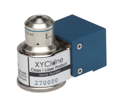
LYKOS®
For use in Human Clinical IVF procedures. Hamilton Thorne offers the LYKOS, & LYKOS® DTS (dynamic targeting system) moveable laser.
View Product
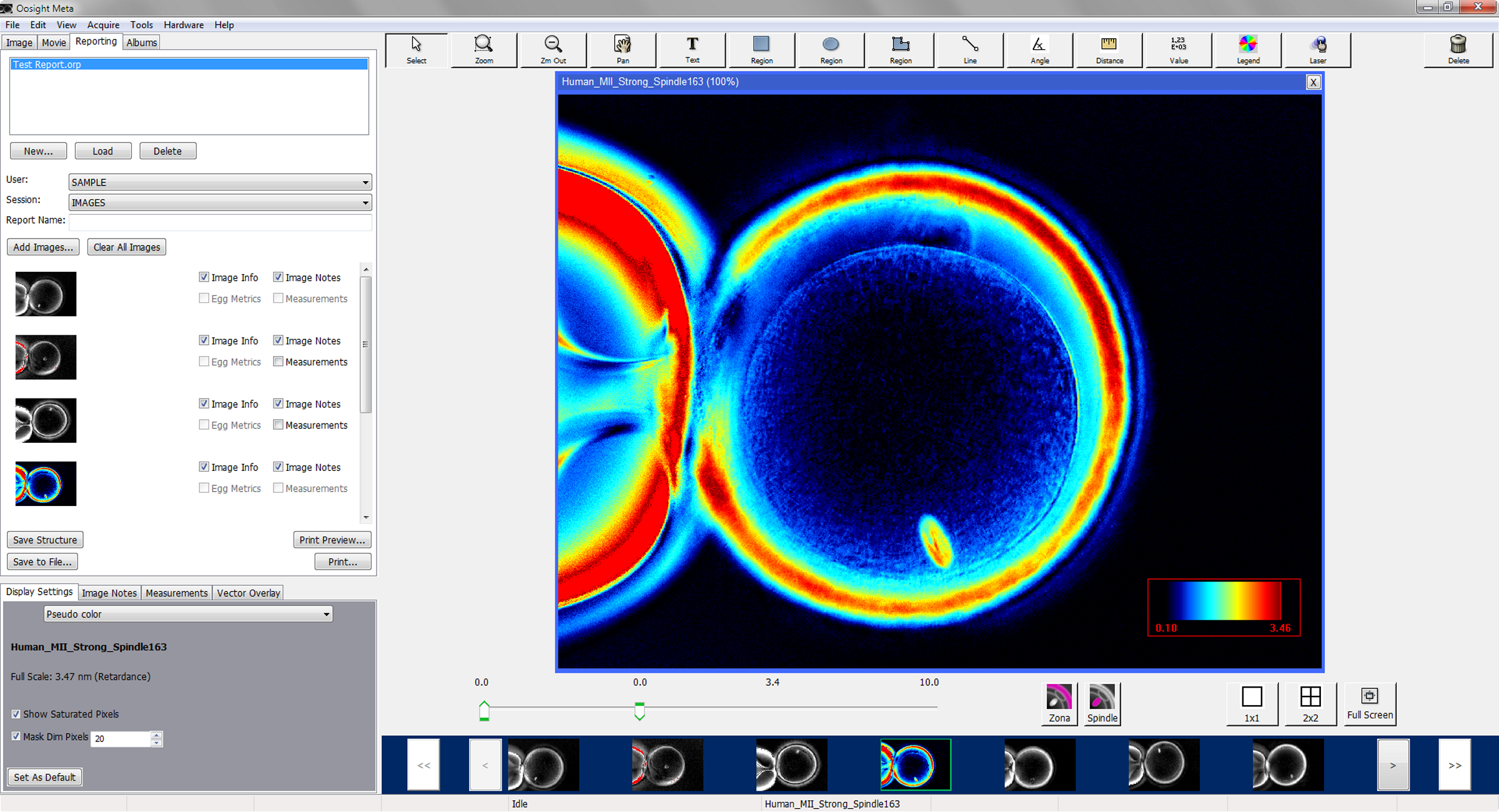
The Oosight Imaging System provides high-contrast live images of the oocyte and spindle to determine which subpopulations are at high risk for producing chromosomally abnormal embryos and can prevent potentially damaging effects from injecting immature oocytes.
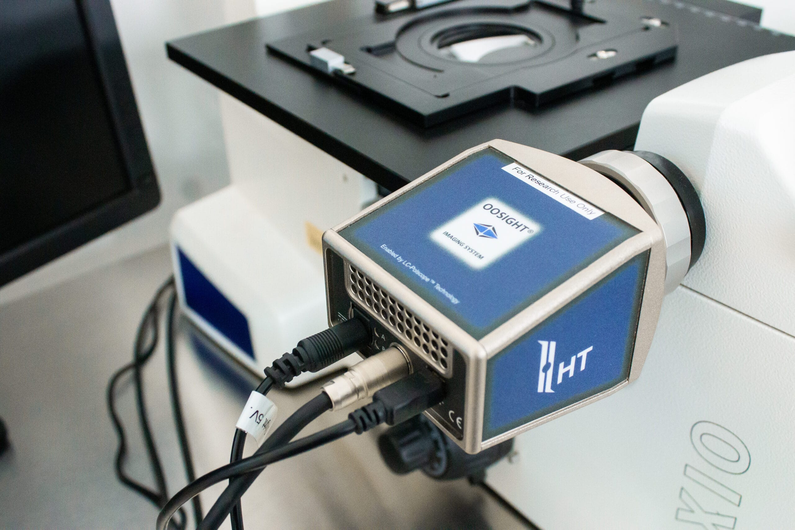
Our unique and patented solid-state, liquid crystal technology is an easy add-on to your ICSI workstation. Oosight software runs on your computer to capture, display, and analyze your images.
Snap and click to report data – it’s really that simple!
Oosight Imaging System is for research purposes only in certain territories.
| Components | Oosight CCD Camera, LC Compensator Optic, Circular Polarizer/Interference Filter, C-Mount Adapter, Power Supply |
| Wavelength of Operation | 546nm |
| Spatial Resolution | Diffraction limited |
| C-Mount | Demagnification C-mount within the range of 0.6x – 0.7x |
| Power Source | 5V 3A with universal input voltage adapters |
| Image Output Format | TIFF |
| CCD Camera | Sensor Size 1/2-inch optical format Image Size 1024 x 1392 pixels Pixel Dimensions 4.65 x 4.65 μm Digital Output 8-bit Binning Modes 1×1, 2×2 |
| Microscope Compatibility | Oosight systems are compatible with many research-grade microscopes, including those made with Leica®, Nikon®, Olympus®, and Zeiss®. Contact us for more information! |
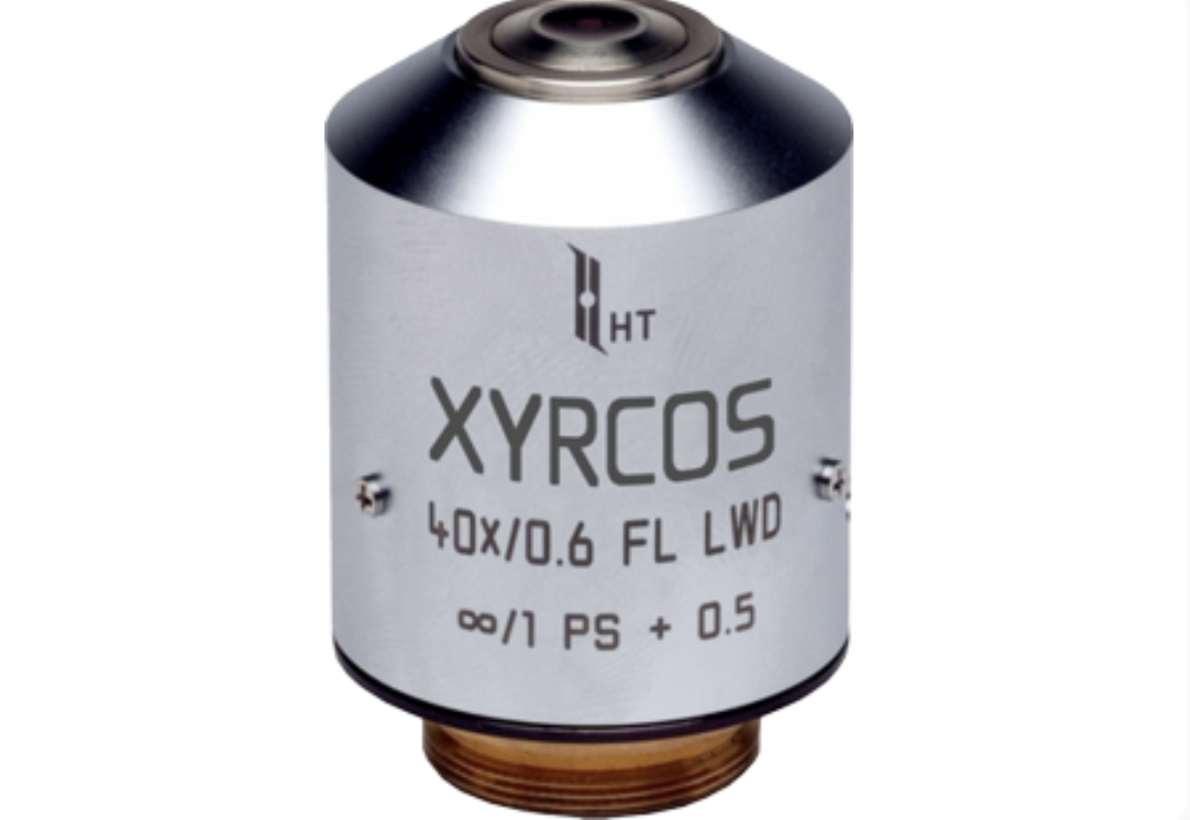
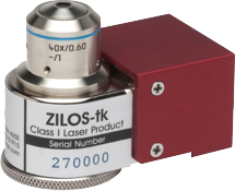
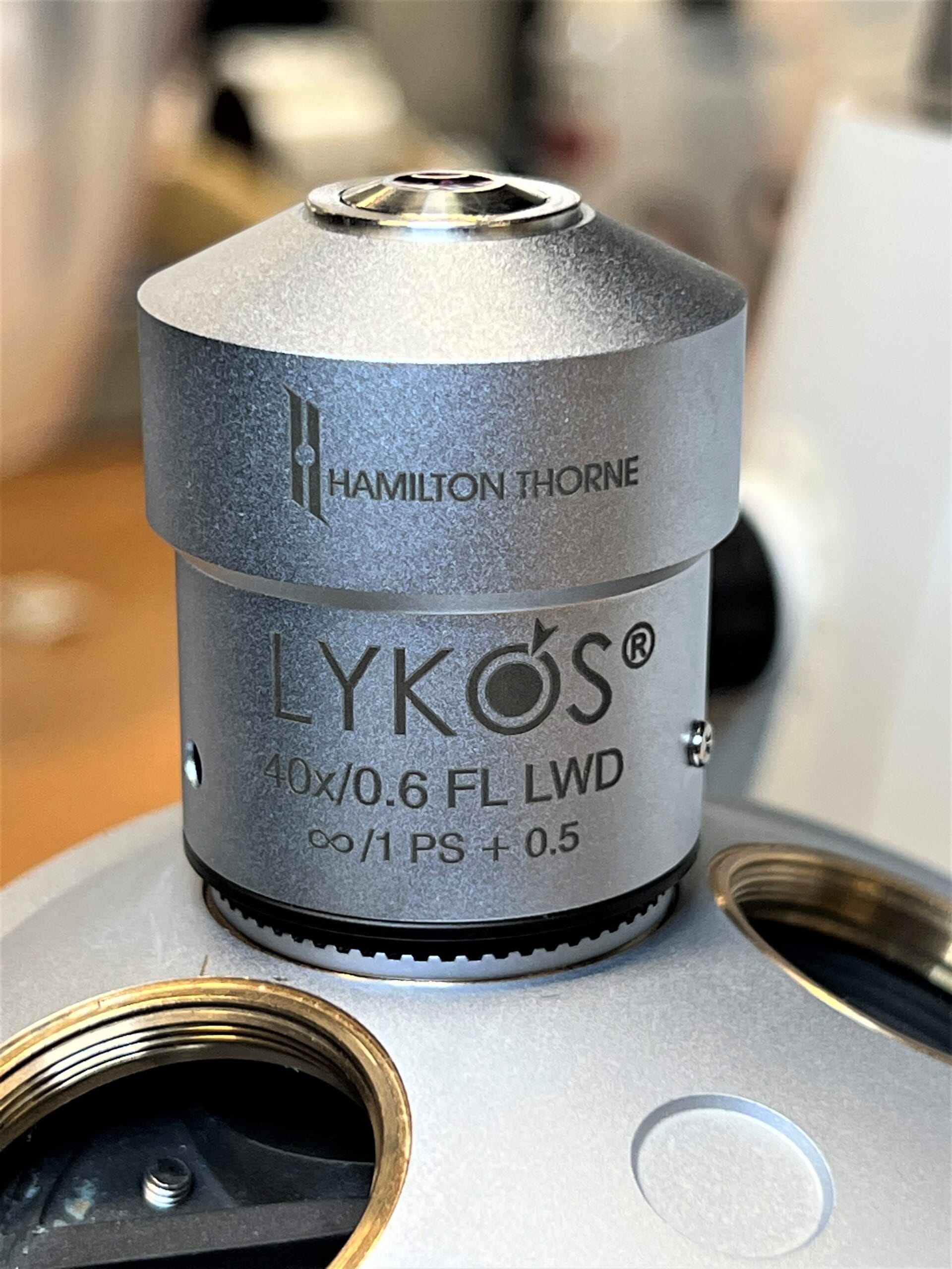
| Laser Compatibility | Hamilton Thorne: XYRCOS, XYClone, LYKOS, ZILOS-tk |
| Environmental Requirements | Operating requirements: Indoor Operating temperature: 15 ˚C to 40 ˚C Operating humidity: 65%, non-condensing Operating altitude: 2000 meters Storage temperature: -10 ˚C to 55 ˚C Storage humidity: 100%, non-condensing Pollution degree: 2 |
| Computer Requirements | Recommended | Minimum |
|---|---|---|
| PC Desktop/Laptop | i5, i7 | Intel Pentium, 2 GHz |
| Operating System | Windows 10, 32- or 64-bit | Windows 7, 32-bit |
| Memory | 4+ GB | 1 GB |
| Hard Disk | 250+ GB | 80 GB |
| Display | 1920×1080, 1920×1200 | 1280 x 1024 |
| USB Ports | USB 2.0 2 available ports | USB 2.0, 2 available ports |
Understanding the oocyte is critical to understanding embryogenesis, and studies show that an oocyte’s disrupted spindle apparatus or a weakened zona pellucida can yield lower pregnancy rates. In fact, studies show that pregnancy is up to 8 times more likely when the inner zona pellucida is well-ordered.1
Adding Oosight to your lab can improve oocyte quality grading success by providing a quantitative and reproducible method to measure biological disruption within the oocyte. The Oosight provides measurements of molecular order and alignment of birefringent structures including: the spindle, zona pellucida, and oolemma.
Improve the efficiency of cryopreservation
The Oosight helps verify vital structural bodies in the oocytes are intact after undergoing freezing procedures.
Nuclear Transfer (SCNT)- No Staining Needed
With Oosight, you can remove the spindle in nuclear transfer techniques without using Hoechst dye DNA staining and minimize damage to vital cytoplasmic organelles.
1. Shen Y, et. al. High magnitude of light retardation by the zona pellucida is associated with conception cycles. Human Reproduction, 2005 Jun; 20(6):1596-606
Oosight can help identify oocytes that may have been falsely labeled MII due to conventional imaging techniques. The system can also help screen for oocytes with highly disrupted spindles, such as those that are multi-polar.
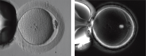
In a conventional contrast image (left) of a human MII oocyte taken just prior to ICSI, structures such as the spindle and multiple layers of the zona pellucida remain invisible. In an Oosight image (right) the spindle is clearly seen to be nicely barrel shaped and the three layers of the zona pellucida are all visible.
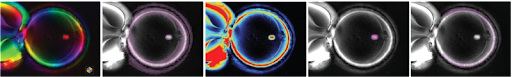
Oosight software visualization tools include (left to right): slow-axis orientation color map, orientation vector overlay, retardance color map, automated SpindleFinder™, and automated ZonaFinder™.
With unprecedented resolution and calibrated setup, Oosight software helps improve your grading routine with reproducibility and the speed needed for micromanipulation.
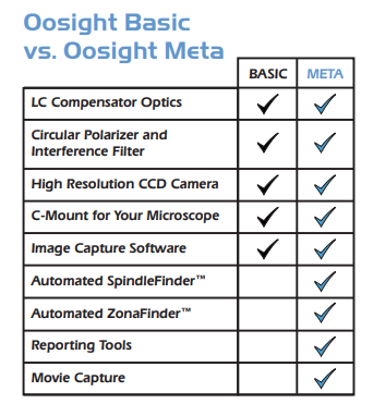
Choose between the Oosight Basic vs Oosight Meta system.
Oosight Basic
The Oosight Basic imaging system combines patented optical, electronic and software elements to produce both live and still images of critical structures within the oocyte, including the meiotic spindle and tri-laminar zona pellucida.
Oosight Meta
The Oosight Meta imaging system combines the image capture capability with additional analysis tools such as automated SpindleFinder™, automated ZonaFinder™, reporting, and Movie Capture to make quantitative, reproducible oocyte analysis possible.
Enter your information below to view this resource.
"*" indicates required fields
Sign in to the distributor portal to gain access to all gated assets and exclusive content.
Case Studies